Daniel Adibi ‘26
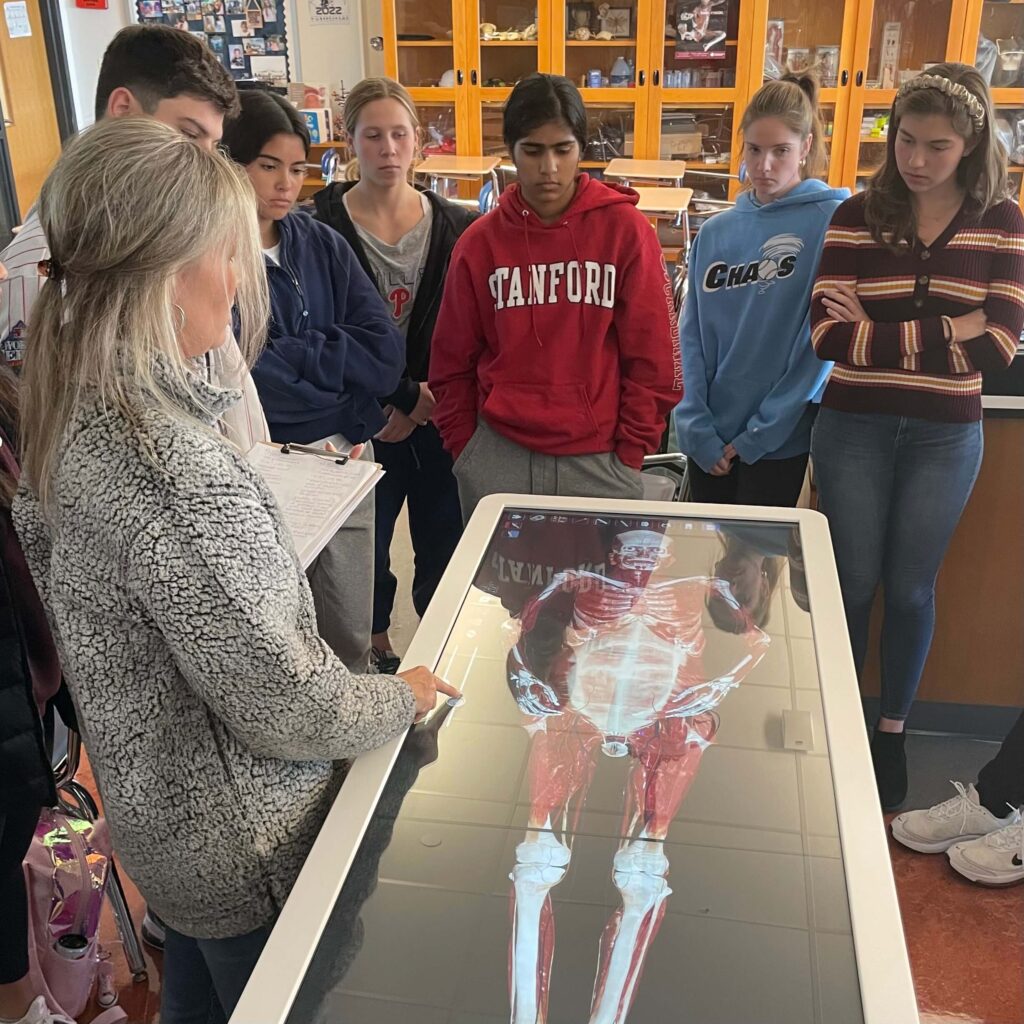
In early October, EA received the Anatomage Table, a new tool capable of rendering virtual anatomical procedures without a physical cadaver. The Clare Foundation, a foundation run by the Clare family which seeks to support EA’s STEM programs, donated the table.
Discussing the Clare Foundation’s donation, Head of School Dr. T.J. Locke states, “[The Clare Foundation] cares about moving STEM projects forward and loves EA. [The foundation] is such a selfless-minded group, and they have previously funded EA’s STEM speaker series. They wanted to do something else to advance the school, so when they heard about the anatomy table idea, they ran with it.”
Thomas Cork, Upper School Science Department Chair, envisions that the Anatomage Table will help teachers introduce new anatomical concepts in a different way. “[The table] basically has stored micro slices of the human body, so imagine that they took a human body, sliced it into many different pieces, and digitized it. It’s crazy to think about how much work that took and how much data is stored – it’s incredible,” explains Cork.
Biology and anatomy teachers aim to use the table to facilitate the teaching of material and to foster interactive learning. Jennifer Jones, Upper School AP Biology and Honors Anatomy and Physiology teacher, remarks, “The table is a great complement to the courses and provides a lot of hands-on learning for the students. The curriculum will not be centered around the table, but [it] will be used to supplement the material being [taught].”

Photo courtesy of EA Communications
Some students have already seen how the table can be integrated to enhance the learning process and understanding of material. Michelle Jiang ‘23, comments on her experience with the table in her AP Biology class, saying, “I remember being able to see the cell structure of various organisms from different environments and thought that was really neat. I am looking forward to using it more during class and I think it’s a great visual tool for learning.”
In addition to providing an in-depth view of subcellular components, the table can be applied to perform a variety of functions – from virtual labs to in-depth explorations of complex anatomical structures. Explaining some specific uses of the table within the curriculum, Jones says, “[Our class] will be starting joints soon. I plan on introducing the various types of joints and will use the table to zoom in to see the structure of the different types of joints on the cadavers … the students can visualize the type of movement that the joint can do as well as what muscles are being moved.” She continues, “Students can also do different surgeries on the cadaver – a catheterization for example. There’s also a dissection tool, so students can dissect through the cadaver as they would with a real cadaver.”
Will Esterhai ‘24, a student in Jones’ Honors Anatomy class, agrees with Jones that the table is a useful resource for visualizing cadavers. He comments, “The Anatomage Table has been an awesome way for us to see the human body up close. It has been a useful study tool and makes memorizing the body fun.”
In addition to viewing cadavers, the table displays other aspects of biology and anatomy. “[Students can] visualize things like blood flow, neural networks, and what the heart is doing while an EKG is being performed. There are high-resolution images of X-rays, MRIs, and CAT scans, and there are over 1300 case studies loaded with [conditions] like arthritis, gunshot wounds, and fractures from a car crash,” Jones adds.
Echoing Jones’ statements about the several capabilities of the table, Adamo Di Carlo ‘24, a student in her AP Biology class, says, “[I] used it once to look at an animal cell. It offered 3D renditions of almost any anatomical and biological aspect of the human body.”
Reiterating the value of the Anatomage Table in EA science classes, Locke concludes, “[The Anatomage Table] is going to be a great teaching tool, which is of course the most important thing, but it’s also a showpiece that demonstrates how much EA cares for science.”